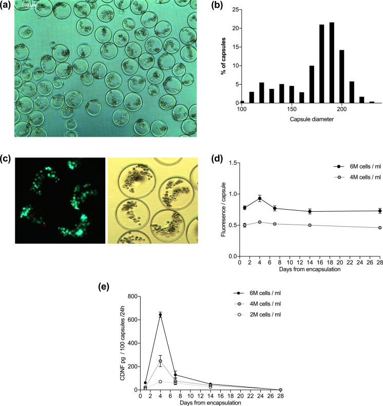
Figure 2.
Microencapsulated wtCDNF ARPE-19 cell clones are viable but the secretion of CDNF decreases with time. (a) APA-microcapsules with cell density of 2 million cells/ml of alginate. Scale bar 100 μm. (b) A density histogram showing the size distribution of the prepared microcapsules. Mean capsule diameter for all the produced capsules was 169 ± 26 μm (range 99–222 μm). (c) Cells stained with LIVE/DEAD kit. Most of the encapsulated cells stained with calcein (live, green), and only a few with ethidium homodimer-1 (dead, red), implying that the cells survived the encapsulation procedure well. (d) Viability test using resazurin solution (Alamar Blue reagent) shows steady metabolic function of the encapsulated cells. (e) wtCDNF release measured in the conditioned media of encapsulated cells analyzed on CDNF ELISA.

