FIGURE 2.

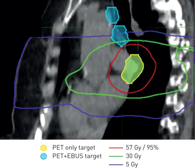
Sagittal view illustrating dose distribution with endobronchial ultrasound (EBUS)-proven malignancy (blue) receiving negligible doses of radiation; positron emission tomography (PET)-positive disease (yellow) received a high dose (>57 Gy).

Sagittal view illustrating dose distribution with endobronchial ultrasound (EBUS)-proven malignancy (blue) receiving negligible doses of radiation; positron emission tomography (PET)-positive disease (yellow) received a high dose (>57 Gy).