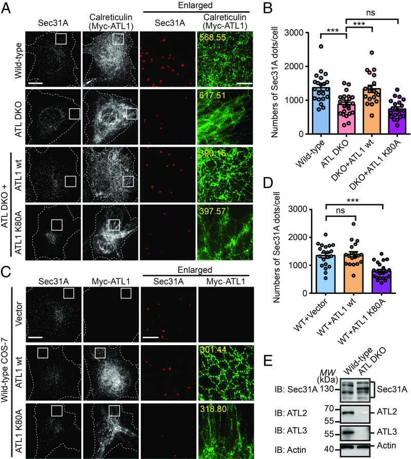
Fig. 1.
Altered COPII pattern in ATL-deleted cells. (A) Nontreated WT, ATL DKO, and Myc-ATL1 WT or Myc-ATL1 K80A transfected ATL DKO COS-7 cells were fixed and stained using anti-Sec31A, anti-calreticulin, or anti-Myc antibodies and imaged using structured illumination microscopy (SIM). Images are projections of 3D datasets (5 μm in z). Dashed lines indicate cell boundaries. Yellow numbers indicate the intensities of green fluorescence (ER marker) inside the white square in pixel (Scale bar, 5 μm or 1 μm in enlarged views). (B) Quantification of the number of Sec31A-labeled structures in A based on SIM (WT, n = 21; ATL DKO, n = 23; ATL DKO cells transfected with Myc-ATL1 WT, n = 19; with Myc-ATL1 K80A, n = 20). Data are presented as mean ± SEM. ***P < 0.001 by 2-tailed Student’s t test; ns, not significant. (C) Wild-type COS-7 cells were transfected with vector, Myc-ATL1 WT or Myc-ATL1 K80A. Twenty-four hours after transfection, cells were fixed and stained using anti-Myc and anti-Sec31A antibodies. SIM images are shown. Yellow numbers indicate the intensities of green fluorescence (ER marker) inside the white square in pixel (Scale bar, 5 μm or 1 μm in enlarged views). (D) Quantification of the number of Sec31A-labeled structures in C based on SIM (for cells transfected with vector, n = 20; with Myc-ATL1 WT, n = 19; with Myc-ATL1 K80A, n = 22). Data are presented as mean ± SEM. ***P < 0.001 by 2-tailed Student’s t test; ns, not significant. (E) Cell lysates from wild-type and ATL DKO COS-7 cells were analyzed by immunoblotting using antibodies against Sec31A, ATL2, or ATL3. Actin is used as a loading control. MW, molecular weight (in all figures).

