Fig. 1.
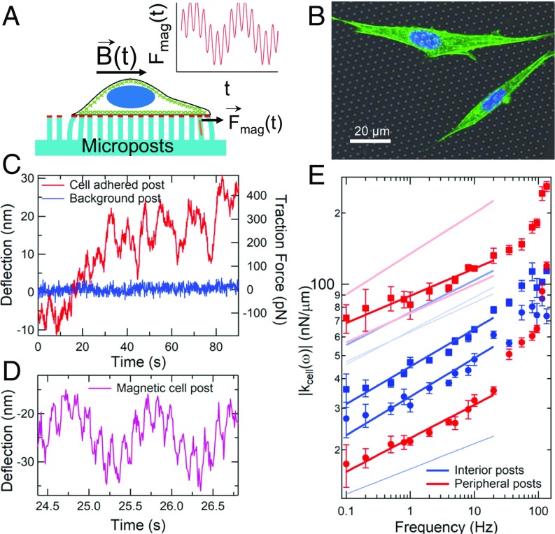
Microposts can be used to measure cytoskeletal dynamics and viscoelasticity. (A) Cell on a flexible micropost array; micropost tips are coupled to the actomyosin cytoskeleton (green) via fibronectin (red). Posts with magnetic nanowires (orange) exert a double-sinusoidal force Fmag(t). (B) Composite white-light image of post array with fluorescence of 3T3 fibroblasts immunostained for actin (green) and nuclei (blue). Actin stress fibers are concentrated near the cells’ peripheries. (C) Typical displacements of a cell-adhered (red) and a nonadhered (blue) post. (D) Deflection of a cortex-adhered magnetic post, driven at 1 Hz and 7 Hz. (E) The computed local cellular rheology is a weak power law function of frequency (symbols); peripheral posts are in red, interior posts are in blue. As shown in Fig. 2, peripheral posts are associated with stress fibers, and interior posts are associated with the cortex. Error bars were determined as described in SI Appendix, Methods. Solid lines are fits of the form |kcell(ω)| = Aωβ with βinterior = 0.13 ± 0.02 (n = 6) and βperipheral = 0.12 ± 0.02 (n = 4) (±SE for each case). Fits for additional magnetic microposts (data points omitted for clarity) are shown as pale lines. See SI Appendix, Fig. S2 for full dataset.

