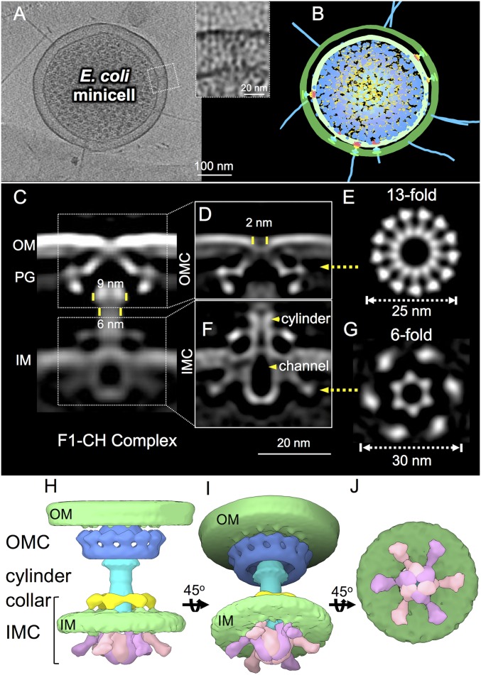
Fig. 1.
In situ structure of the E. coli F-plasmid type IV secretion machine revealed by CryoET and subtomogram averaging. (A) A tomographic slice from a representative E. coli minicell showing multiple T4SS machines embedded in the cell envelope; the boxed region was magnified to show an F1-CH complex. (B) A 3D surface view of the E. coli minicell in A showing T4SS machines and F pili. (C) A central slice of the averaged structure of the F1-CH complex in the cell envelope. Diameters of the flare and cylinder are shown. (D) After refinement, details of the OMC are visible, including a 2-nm gap in the OM. (E) A cross-section view of the region in D marked by a yellow arrow shows 13-fold symmetry of the OMC. (F) After refinement, details of the IMC are visible, including a periplasmic collar, cylinder and central channel (yellow arrowheads), and cytoplasmic inverted “V” structures. (G) A cross-section view of the region in F marked by a yellow arrow shows sixfold symmetry of the IMC. (H–J) Three-dimensional surface renderings of the F1-CH complex shown in different views.

