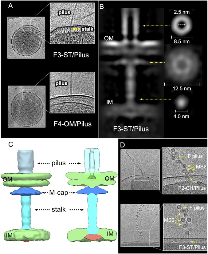
Fig. 4.
In situ structures of F-encoded F3-ST/Pilus and F4-OM/Pilus complexes. (A) Tomographic slices from representative E. coli minicells showing the F pilus associated with an envelope-spanning stalk (F3-ST/Pilus) or the OM in the absence of an associated periplasmic density (F4-OM/Pilus). (B) A central slice of the averaged structure of the F3-ST/Pilus complex and cross views of the pilus, “mushroom-cap,” and stalk showing the absence of a central channel; cross-sections views and dimensions are presented for the various structures at positions indicated by yellow arrows. (C) Three-dimensional surface rendering of the F3-ST/Pilus complex and cutaway view with architectural features indicated. (D) MS2 bacteriophage binds to the sides of pOX38-encoded F pili docked onto F2 and F3 basal structures elaborated by intact E. coli cells.

