Figure 8.

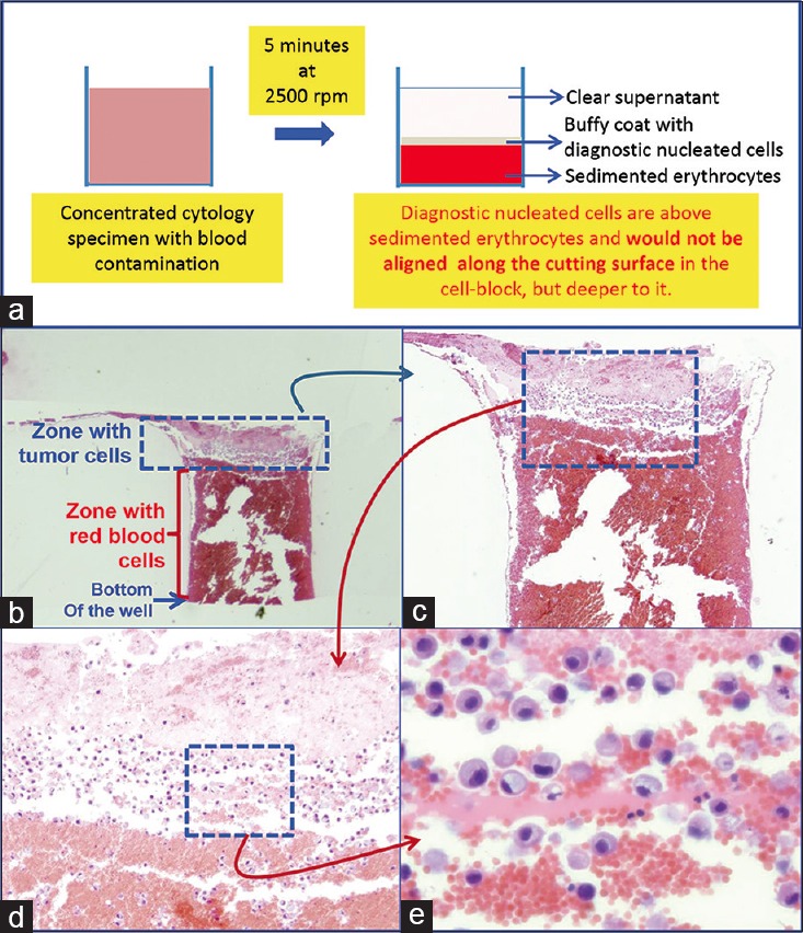
Blood contaminated cytology specimen (H and E). (a) Schematic showing result of centrifuging the blood-rich concentrated specimen with diagnostic cells which group with nucleated cells in the buffy coat area above red blood cells. (b) The longitudinal sections of one of the wells in the cell-block made with Nano NextGen CelBloking™ kit. (c) The bottom of the wells is predominantly red blood cells with tumor cells on the top which will be way deep to the actual cutting surface of usual cell blocks (H and E). (d and e) Higher magnification showing the diagnostic tumor cells in the area corresponding with the buffy coat (H and E)
