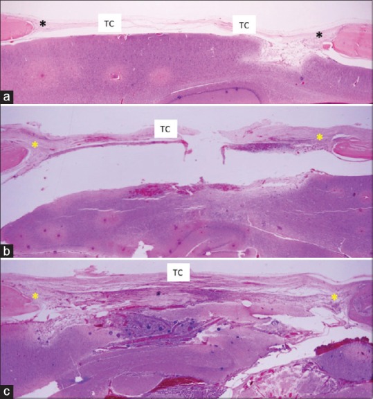
Figure 2.

Histological section of 30-day experimental groups: panoramic image (H and E, ×2.5). (a) Control group showing presence of bony defect filled by a thin layer of connective tissue, mainly in the central region; (b) Group without coaptation: bony defect filled by a thicker layer of connective tissue, mainly within the central region; (c) Coaptation group: bony defect filled by connective tissue with greater organization in the central region. *Bony defect; TC – Connective tissue
