Fig. 1.
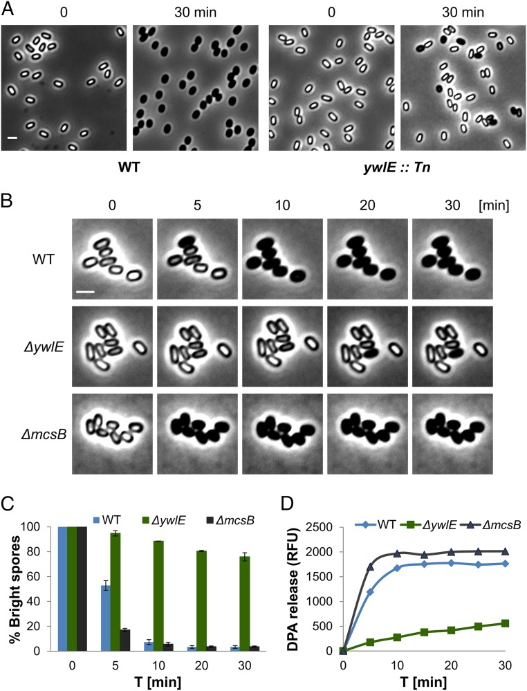
Spore germination is facilitated by Arg dephosphorylation. (A) Spores of PY79 [wild type (WT)] and ywlE::Tn were incubated with l-Ala (10 mM) and observed by light microscopy before (t = 0) and after (t = 30 min) l-Ala addition. Shown are phase contrast images from a representative experiment out of three independent biological repeats. (Scale bar: 1 μm.) (B) Spores of PY79 (WT), BZ16 (∆ywlE), and BZ129 (∆mcsB) strains were incubated on agarose supplemented with l-Ala (10 mM) and monitored by time lapse microscopy. Shown are phase contrast images from a representative experiment out of three independent biological repeats. (Scale bar: 1 μm.) (C) Spores of PY79 (WT), BZ16 (∆ywlE), and BZ129 (∆mcsB) strains were incubated on agarose supplemented with l-Ala (10 mM) and monitored by time lapse microscopy. Data are presented as percentages of the initial number of the phase-bright spores. Shown are average values and SDs obtained from three independent biological repeats (n ≥ 300 for each strain). (D) Spores of PY79 (WT), BZ16 (∆ywlE), and BZ129 (∆mcsB) strains were incubated with l-Ala to trigger germination. DPA release to the medium was determined by Tb-DPA assay. Presented are relative fluorescence units (RFUs) measured at 545 nm with excitation at 270 nm. Shown is a representative experiment out of three independent biological repeats.

