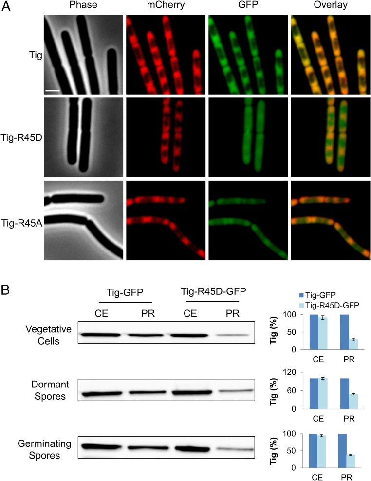
Fig. 3.
Tig-ribosome association is dependent on Arg dephosphorylation. (A) BZ118 (tig-gfp, rpsB-mCherry), BZ119 (tig-R45D-gfp, rpsB-mCherry), and BZ120 (tig-R45A-gfp, rpsB-mCherry) strains were grown to a mid logarithmic phase in LB at 37 °C and visualized by fluorescence microscopy. Shown are images of phase contrast (Phase), fluorescence from mCherry (red), and GFP (green), and an overlay of red and green fluorescence from a representative experiment out of three independent biological repeats. (Scale bar: 1 μm.) (B) Intact ribosomes were purified from CEs of the vegetative cells, dormant and germinating spores of BZ118 (tig-gfp, rpsB-mCherry) and BZ119 (tig-R45D-gfp, rpsB-mCherry) strains. Germination was induced by suspending the spores in 10 mM l-Ala for 10 min. Western blot analysis was carried out using an antibody against GFP, detecting the levels of Tig and TigR45D in CEs and in PRs (Left). The signal from GFP fusion proteins was quantified by MetaMorph software (version 7.7, Molecular Devices) (Right). The signal from Tig-GFP was used to normalize Tig expression levels in CEs and PRs, separately. Shown are average values and SDs obtained from two independent biological repeats.

