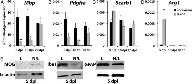
Fig. 4.
Differential mRNA and protein expression in NR-labeled lesion. NR-labeled lesion and adjacent nonlesion tissue at 5, 10, and 20 dpl were dissected out and processed for RT-qPCR (A–D) or protein (E) analyses (n = 6–8 animals per time point examined). The expression of oligodendrocyte-specific genes, Mbp (A) and Pdgfrα (B), was normalized to B2m housekeeping gene; macrophage-specific Scarb1 (C) and Arg1 (D) mRNA levels were normalized to Ppia. (E) The amount of myelin oligodendrocyte glycoprotein (MOG), macrophage/microglia marker Iba1, and astrocyte marker GFAP in NR-labeled lesion (L) and unlabeled nonlesion (N/L) tissue at 5 dpl detected by Western blot. β-actin was used for loading control. Data presented as mean ± SEM. *P < 0.05, **P < 0.01.

