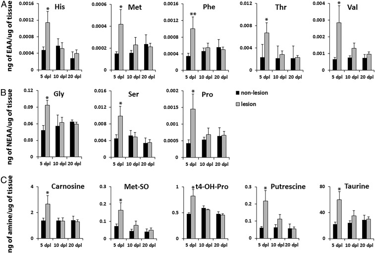
Fig. 6.
Mass spectrometry generated metabolomics of the lesion environment during remyelination. NR-labeled lesion and adjacent unlabeled nonlesion tissue (n = 6–9 animals per group) were dissected out at 5, 10, and 20 dpl and processed for mass spectrometry of 21 AAs and 21 biogenic amines, using ultraperformance liquid chromatography electrospray ionization–tandem mass spectrometry analysis. Five essential AAs (A), 3 nonessential AAs (B), and 5 biogenic amines (C) were significantly increased in NR lesion compared with nonlesioned tissue at 5 dpl during active inflammation. All metabolite AAs and metabolite levels in NR lesion were reduced to control levels at 10 and 20 dpl during remyelination. Data are presented as mean ± SEM. **P < 0.01, *P < 0.05.

