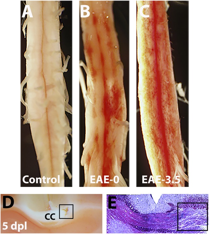
Fig. 7.
NR labels demyelinating lesion in the brain and EAE. (A–C) Representative images showing NR labeling of spinal cord from control mice without EAE (A), or with EAE at clinical score 0.0 (B) and 3.5 (C). Note that NR is not detectable in mice without EAE (control) but is detected in mice with EAE before clinical deficit. (D and E) NR labels lysolecithin-induced lesion (square) in the corpus callosum at 5 dpl. Bright field images showing NR (D) and toluidine blue staining (E) of demyelinated area.

