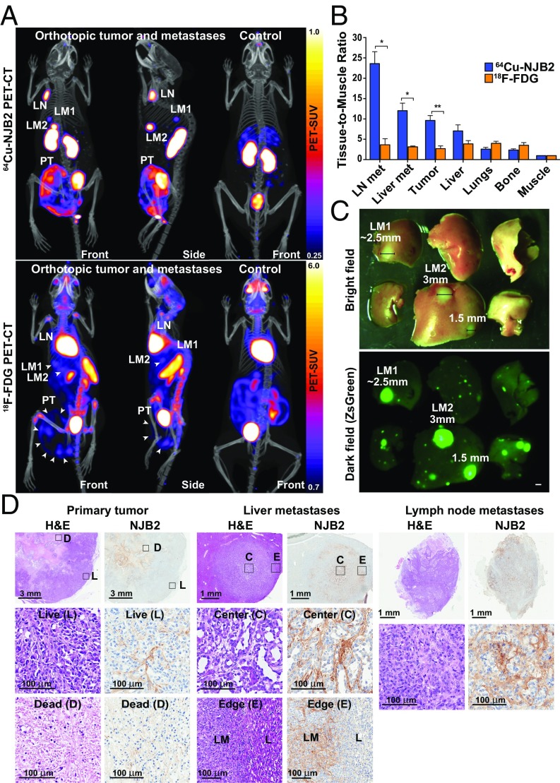
Fig. 3.
NJB2 nanobody detects discrete metastases to liver and LNs with a higher signal-to-noise ratio compared with 18F-FDG PET/CT imaging. (A) NSG mice were injected in the mammary fat pad with 0.25 × 10⁶ LM2 cells. The tumors were allowed to grow and metastasize for 6 wk. Mice were imaged with 18F-FDG or 64Cu-NJB2. The 64Cu-NJB2 PET/CT images were compared with 18F-FDG images of the same mouse. Representative PET/CT images (front and side views) imaged with 64Cu-NJB2 reveal primary tumor (PT), discrete LN, and liver metastases (LM1, LM2). In 18F-FDG PET/CT, signals were observed in the primary tumor (PT), heart, kidney, spleen, bladder, and Harderian glands. Movies S4–S7 show 3D visualization. (B) In vivo PET signals were quantified 2 h after injection of 64Cu-NJB2 and 90 min after injection of 18F-FDG. 64Cu–NJB2 was able to detect primary tumors and metastases with a significantly higher TMR compared with 18F-FDG PET/CT. Data were analyzed by two-tailed unpaired t test. (n = 3 for mice with orthotopic tumors; SI Appendix, Fig. S4). (C) Bright-field and fluorescence microscopy of resected liver lobes with metastases (SI Appendix, Fig. S5). (Scale bar, 1 mm.) (D) H&E staining and IHC with NJB2 of resected organs of mice imaged in A. EIIIB was observed in the ECM surrounding live tumor cells (marked as “L”) but not in the dead/necrotic regions (“D”). Strong EIIIB staining was observed in the ECM of liver metastases (LM) and LN Met but not in the ECM of normal liver (“L”).

