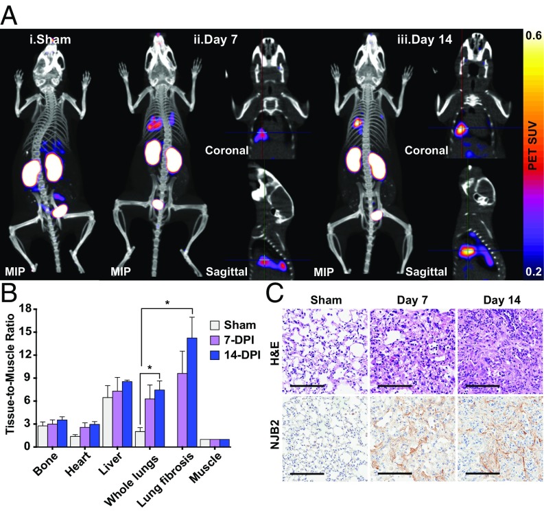
Fig. 5.
NJB2 detects pulmonary fibrosis in a bleomycin-induced lung fibrosis model. (A) Pulmonary fibrosis was induced by a single intratracheal administration of 0.035U of bleomycin sulfate into C57BL/6 mice. Sham and bleomycin-treated mice (7 and 14 d after bleomycin administration) were imaged with 64Cu-NJB2 PET/CT. Representative PET/CT images of mice that were sham-treated (i), 7 d after bleomycin treatment (ii), and 14 d after bleomycin treatment (iii). Fibrotic lesions were visible in mice at 7 and 14 d postadministration. The cross-hairs in the coronal and sagittal slices mark the locations of the fibrotic lesions within the lungs. Movies S14–S22 show 3D visualization (SI Appendix, Fig. S7A). MIP, maximum intensity projection. (B) The in vivo PET signals were quantified, and the TMR was significantly higher in bleomycin-treated mice at day 14 compared with sham controls (analyzed by two-tailed unpaired t-test). (C) Following 64Cu-NJB2 PET/CT imaging, lungs were resected for histopathological analysis. Representative H&E images of lung sections indicated bleomycin-induced pulmonary fibrosis progression in mice at days 7 and 14 after administration (SI Appendix, Fig. S7B). No fibrosis was observed in sham-treated mice. (Scale bars, 100 μm.)

