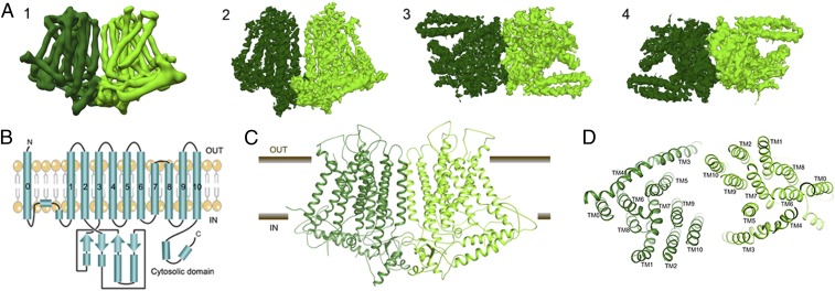
Fig. 1.
Cryo-EM structure of the OsOSCA1.2 protein. (A, Left to Right) (1) Parallel to membrane plane view of unsharpened cryo-EM density map used for initial chain tracing and (2–4) sharpened 4.9-Å map used for model building and refinement [membrane plane view (2), extracellular view (3), and intracellular view (4)]. (B) Protein topology of OsOSCA1.2. The OsOSCA1.2 model is shown in the TM plane (C) and from the extracellular side (D).

