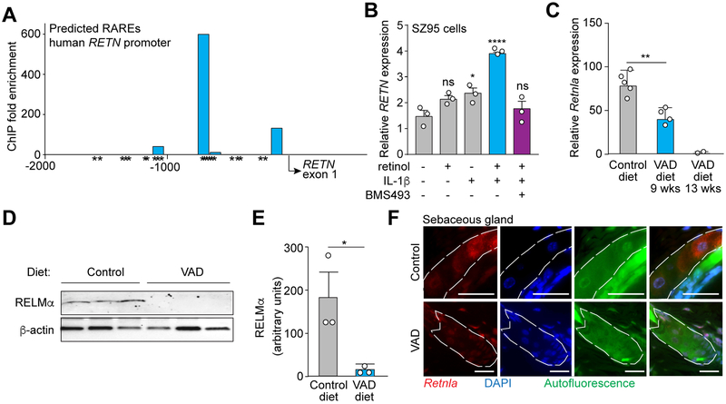
Figure 5: Vitamin A is required for RELMα expression.
(A) RAR binding to the human RETN promoter was measured by chromatin immunoprecipitation (ChIP) with an anti-RAR antibody. Predicted retinoic acid response elements (RAREs) were identified by in silico analysis using NUBIScan (Podvinec et al., 2002) and are indicated by an asterisk (*). Predicted RARE sequences are shown in Figure S6. Data are representative of two independent experiments.
(B) qRT-PCR analysis of human RETN expression in the human sebocyte cell line SZ95. Cells were treated with retinol, IL-1β, the pan-RAR inhibitor BMS493, or a combination.
(C) Mice on a Vitamin A deprived diet express less Retnla transcript. qRT-PCR analysis of Retnla expression in the skin of mice on a control or vitamin A-deficient (VAD) diet administered for 9 weeks or 13 weeks.
(D,E) Mice on a Vitamin A deprived diet express less RELMα protein. Western blot detection of RELMα in skin of mice fed a control or VAD diet (D), with quantification (E).
(F) FISH detection of Retnla in mouse skin shows decreased Retnla transcripts in sebaceous glands (dashed line) of VAD diet-fed mice. Scale bar, 25 μm. Data are representative of at least two experiments. *P<0.05, **P<0.01,***P<0.001 by One-way ANOVA (B, C); unpaired t-test (E).

