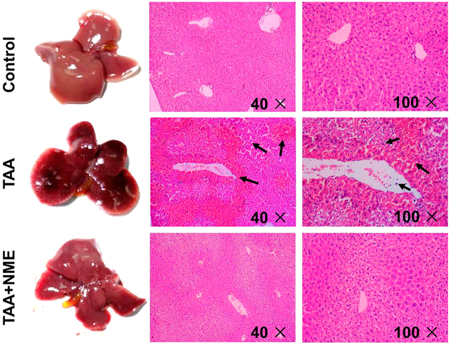
Figure 2.
Histological examination. Livers from mice administered a single dose of TAA showed dark color and larger cholecyst; pretreatment with NME before TAA administration resembles that of normal control. In TAA-treated mouse liver sections, the arrows indicate necrocytosis and neutrophil infiltration. NME treatment protected the histomorphology of liver and reduced inflammatory cells infiltration.

