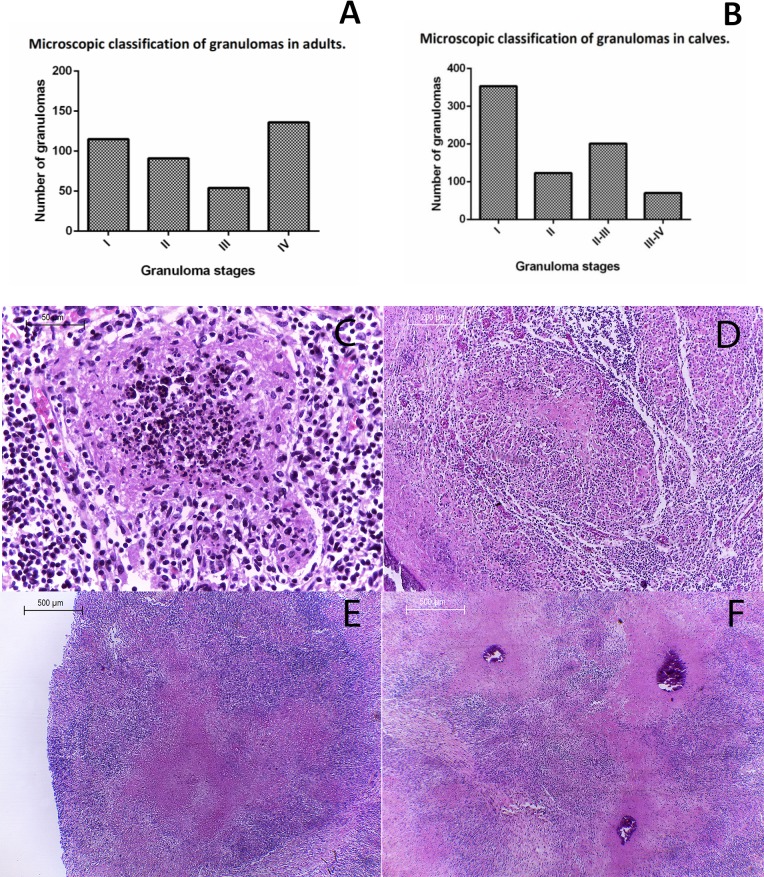
Fig 4. Histopathological classification of granulomas found in lymph nodes from calves four months of age or younger.
(A) Graph of granuloma stages identified in adults, showing a higher frequency of stages I and IV. (B) Graph showing a higher frequency of stage I granulomas in calves. H&E (C) Stage I: 400X, small diffuse foci of infiltrated inflammatory cells, mainly macrophages, epithelioid macrophages and abundant cellular detritus at the center of the lesion. (D) Stage II: 200X, Larger structure with epithelioid macrophages, multinucleated giant cells, cell debris and necrosis. (E) Stage II-III: 40X, large necrotic areas without borders, absence of connective tissue capsule or lymphocyte accumulation in the periphery. (F) Stage III-IV: 40X, lesions showing extensive necrotic areas, mineralization and cell debris without defined borders, surrounded by macrophages, absence of connective tissue capsule or lymphocytes in the periphery; lesions tend to coalesce and fuse.

