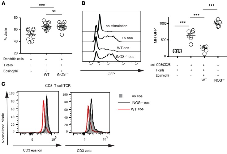
Figure 4. Eosinophils alter TCR signal transduction.
(A) In vitro MLRs established using the coculture of bone marrow–derived BALB/c dendritic cells, and B6 T cells with a 2:1 ratio of E1-polarized B6 eosinophils directly added to the culture. T cell viability was determined flow cytometrically after 5 days of coculture with no, WT, or iNOS–/– eosinophils as described in the graph. Representative data from 1 out of 3 similar experiments. (B) In vitro MLRs established using the coculture of anti–CD3/CD28 Dynabeads with Nur77 T cells and a 2:1 ratio of E1-polarized eosinophils. Nur77-driven GFP expression was used as a gauge of TCR stimulation in CD8+ T cells after 36 hours of coculture with no, WT, or iNOS–/– eosinophils. Representative of 1 out of 3 separate experiments. (C) Evaluation of the TCR/CD3 complex integrity by quantification of TCRβ immunoprecipitations with coassociated CD3ζ or CD3ε on CD8+ T cells isolated from in vitro MLRs in the presence of no eosinophils (gray) versus WT (red line) or iNOS–/– eosinophils (black line). Data representative of 1 out of 5 mice. All statistics performed by Mann-Whitney U test. ***P < 0.001, nsP > 0.05.

