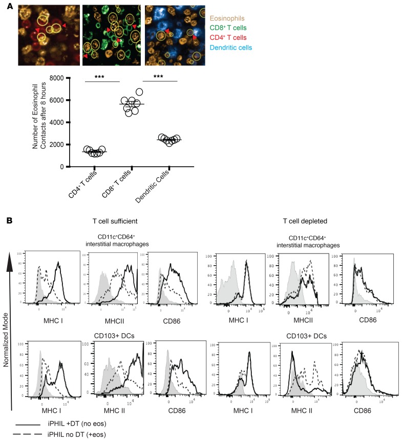
Figure 6. Eosinophil interaction with and phenotypic alteration of myeloid cells.
(A) In vitro MLRs established using the coculture of fluorescently labeled bone marrow–derived BALB/c dendritic cells, and fluorescently labeled CD8+ and CD4+ B6 T cells with a 2:1 ratio of E1-polarized fluorescently labeled B6 eosinophils. Eosinophil–T cell–dendritic cell interactions were analyzed using Harmony Software, with the yellow circles in the top graphic representing interactions. The bottom panel represents the number of eosinophil contacts with CD4+ T cells, CD8+ T cells, or dendritic cells. Data representative of 2 separate experiments. (B) BALB/c lungs were engrafted into B6 iPHIL mice treated with DT (eosinophil deficient) or saline (eosinophil sufficient). The phenotypes of CD11c+CD64+ interstitial macrophages and CD103+ dendritic cells were evaluated flow cytometrically in engrafted lungs in the presence of the full complement of T lymphocytes (left panels) or after depletion of both CD4+ and CD8+ T cells (right panel). Representative of 2 separate sets of transplants. All statistics performed by Mann-Whitney U test. ***P < 0.001.

