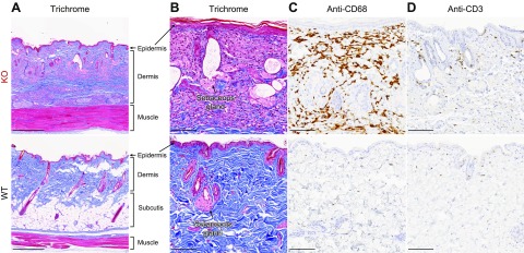
Figure 2.
Inflammation and lipoatrophy are evident in aged Fabry rat skin. Back-skin sections from aged (90 wk old) male rats were stained with Masson’s trichrome (A, B); anti-CD68, a marker of macrophages (C); or anti-CD3, a pan T lymphocyte marker (D). Images are representative of sections from 3 WT and 3 KO male rats. Original scale bars, 500 µm (A) and 100 µm (B–D).

