Figure 3.
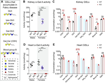
Fabry rat kidney and heart have undetectable α-Gal A activity and accumulate α-galactosyl GSLs. A) List of GSLs with terminal α-galactose that accumulate in Fabry disease. Carbohydrate portions of GSLs are represented using the established symbol nomenclature for glycans. B) α-Gal A activity was measured in 13-wk-old kidney homogenate using a 4-methylumbelliferyl substrate. Biologic replicates include 5 WT males, 5 KO males, 6 WT females, 6 HET females, and 3 KO females. C) Kidney GSL quantification by nanospray ionization-mass spectrometry from 3 WT and 3 KO males at 13 wk (log scale y axis). When GSL levels in KO tissue are statistically elevated above WT, the fold increase is shown above in red. GSLs detected in KO, but not WT, kidneys are highlighted with a blue box. D) Same as in B but α-Gal A activity in heart homogenate. The number of heart biologic replicates is 6 for WT and KO males and WT and HET females and 3 for KO females. E) Same as in C but GSL quantification in heart (log scale y axis). B, D) Male enzyme activity means are compared with an unpaired, 2-tailed t test, and female enzyme activity means (gray box) are compared with a 1-way ANOVA and Dunnett’s multiple comparison test. C, E) WT and KO GSL means are compared with unpaired, 2-tailed t tests. Gal-Gb3, Gb3 with 1 galactose extension; Gal-lyso-Gb3, lyso-Gb3 with 1 galactose extension; Gal2-Gb3, Gb3 with 2 galactose extensions; GalNAc, N-acetylgalactosamine; GlcNAc, N-acetylglucosamine. *P < 0.05, **P < 0.01, ***P < 0.001.

