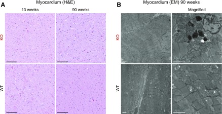
Figure 6.
Myocardium appears similar to WT, but some electron-dense structures are observed in Fabry rat cardiomyocytes. A) H&E-stained myocardium from 13- to 90-wk-old rats. Thirteen-week-old images are representative of n = 3 each: WT males, KO males, WT females, HET females, and KO females. Ninety-week-old images are representative of n = 3 each: WT and KO males. Original scale bars, 100 µm. B) Representative electron microscopy (EM) images of 90-wk-old male myocardium. Original scale bars, 2 µm. Arrows point to inclusions.

