Figure 8.
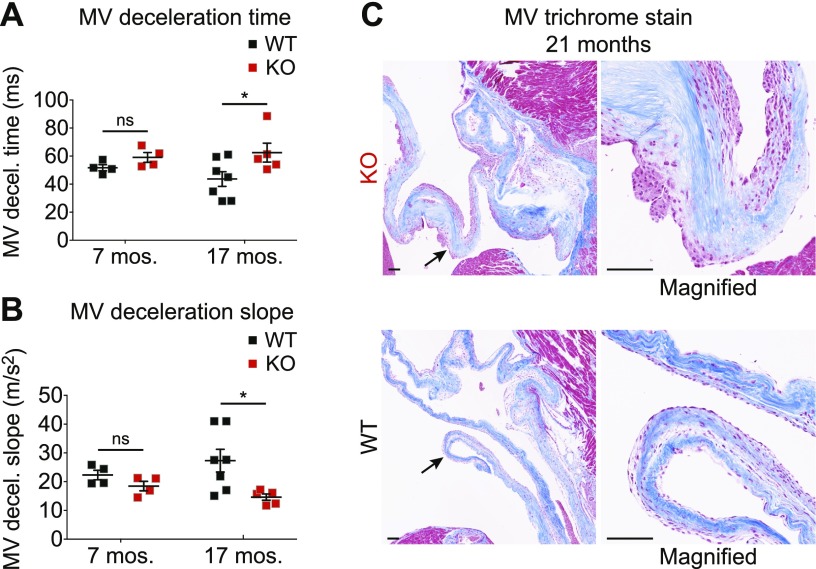
Aged Fabry rats show evidence of mitral valve thickening. A, B) Mitral valve (MV) deceleration time (A) and MV deceleration slope (B) were obtained from echocardiography measurements. Data were obtained from 4 WT and 4 KO male rats at 31 wk (7 mo) and 7 WT and 5 KO male rats at 74–77 wk (17 mo). MV echocardiography data were analyzed using a 2-way ANOVA, and genotype differences were determined with Bonferroni’s multiple comparisons test. C) Ninety-week-old (21 mo) heart sections were stained with Masson’s trichrome, and representative valve images are shown. Arrows point to the section of the valve that is magnified; original scale bars, 100 µm. *P < 0.05.

