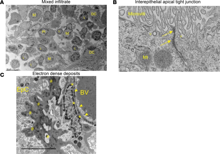
Figure 9. Ultrastructural evaluation reveals extensive leukocyte aggregation and inflammatory changes to the stroma, while maintaining tight junction integrity.
(A) Various cell types were readily identifiable by TEM, including lymphocytes, monocytes, dendritic cells, and plasma cells, confirming the immunofluorescence findings. Scale bar: 10 μm. M, macrophage; Pl, plasma cell. (B) Interepithelial tight junctions found near the apical surface of the CP cells appear normal. Scale bar: 200 nm. Arrows, tight junction; V, vesicle; Mt, mitochondria. (C) The CP stromal space has extensive thickening of the basement membrane and deposition of osmophilic substances, consistent with immune complexes or antibodies. Scale bar: 5 μm. BV, blood vessel; arrowheads, endothelial cells; *, endothelial basement membrane deposition; #, epithelial basement membrane deposition.

