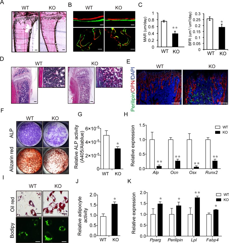
Fig 4. Irx5 is required for osteogenesis and adipogenesis.
(A) von Kossa staining of tibia from 4-week-old WT and Irx5 KO mice. Scale bar, 300 μm. (B–C) Calcein-alizarin red double labeling of tibiae from 4-week-old WT and Irx5 KO mice. Representative images visualized by fluorescent microscopy of periosteal bone and trabecular bone (B). Scale bars, 30 μm (top) and 100 μm (bottom). MAR and BFR (C). Data are presented as mean ± SD, n = 4 in each group. (D) HE staining of tibiae from 4-week-old WT and Irx5 KO mice. Scale bars, 300 μm (left) and 100 μm (right). (E) Immunostaining of Perilipin A/B (green) and OPN (red) of tibiae from 4-week-old WT and Irx5 KO mice. Scale bar, 300 μm. (F) ALP staining of osteoblasts cultured with osteoblast differentiation medium for 7 days, and bone nodule formation visualized by alizarin red staining of osteoblasts cultured for 21 days. (G) Statistical analysis of ALP activity and Alamar Blue activity in the osteoblast differentiation culture via colorimetric readout (A405) and fluorescencent readout, respectively. Data are presented as mean ± SD, n = 4 in each group. (H) Gene expression analysis of osteoblast cultured for 4 days was examined by qRT-PCR. Data are presented as mean ± SD, n = 4 in each group. (I) Oil Red and Bodipy staining of adipocytes cultured with adipocyte differentiation medium for 6 days. Scale bar, 10 μm. (J) Statistical analysis of percentage of Oil Red positive area via Image J. Data are presented as mean ± SD, n = 4 in each group. (K) Gene expression analysis of adipocyte cultured for 6 days was examined by qPCR. Data are presented as mean ± SD, n = 3 in each group. **P < 0.01; *P < 0.05 versus WT. Data associated with this figure can be found in S1 Data. ALP, alkaline phosphatase; BFR, bone formation rate; DAPI, 4ʹ,6-diamidino-2-phenylindole; HE, hematoxylin–eosin; KO, knockout; MAR, mineral apposition rate; OPN, osteopontin; qPCR, quantitative polymerase chain reaction; qRT-PCR, quantitative reverse transcription polymerase chain reaction; WT, wild type.

