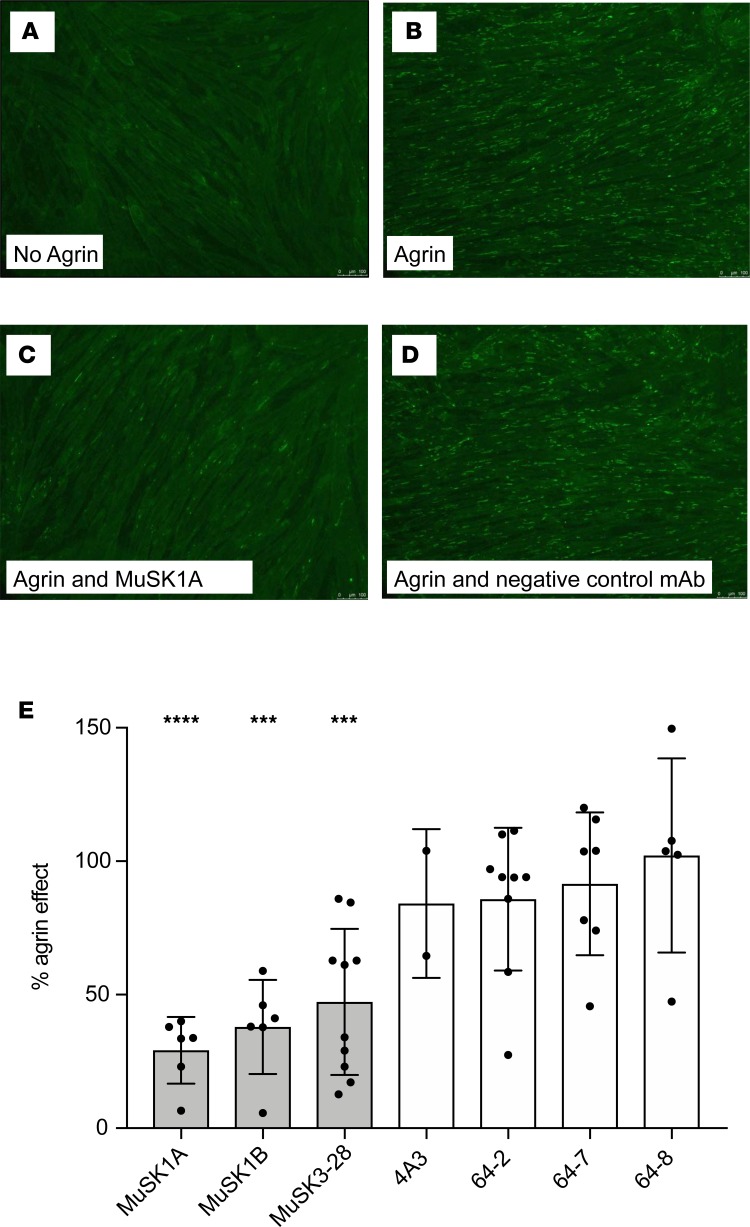
Figure 4. AChR-clustering assay in C2C12 mouse myotubes demonstrates pathogenic capacity of MuSK mAbs.
The presence of agrin in C2C12 myotube cultures leads to dense clustering of AChRs that can be readily visualized with fluorescent α-bungarotoxin and quantified. Pathogenic MuSK autoantibodies disrupt this clustering. Three different human MuSK-specific mAbs, the humanized murine control MuSK mAb 4A3, and 3 human non–MuSK-specific mAbs derived from AChR MG patient plasmablasts (plasmablasts 64-2, 64-7, and 64-8) were tested for their ability to disrupt the AChR clustering. Each mAb was added to the cultures at 1 μg/mL. (A–D) Representative images (original magnification, ×100) from the clustering experiments are shown. (A) Cultured myotubes do not show AChR clustering until (B) agrin is added (bright spots reveal AChR clusters). (C) The mAb MuSK1A added at 1 μg/mL inhibits clustering (D), whereas a control mAb does not inhibit the formation of AChR clusters. (E) Clustering of AChR was quantified relative to the measured effect of agrin. Quantitative results are normalized to clustering induced by only agrin. Each data point represents the mean value from an independent experiment. Bars represent the mean of means and error bars the SDs. Multiple-comparisons ANOVA (against the pooled results for the 3 human non–MuSK-specific mAbs), Dunnett’s test; *P < 0.05, **P < 0.01, ***P < 0.001, and ****P < 0.0001, shown only when significant.

