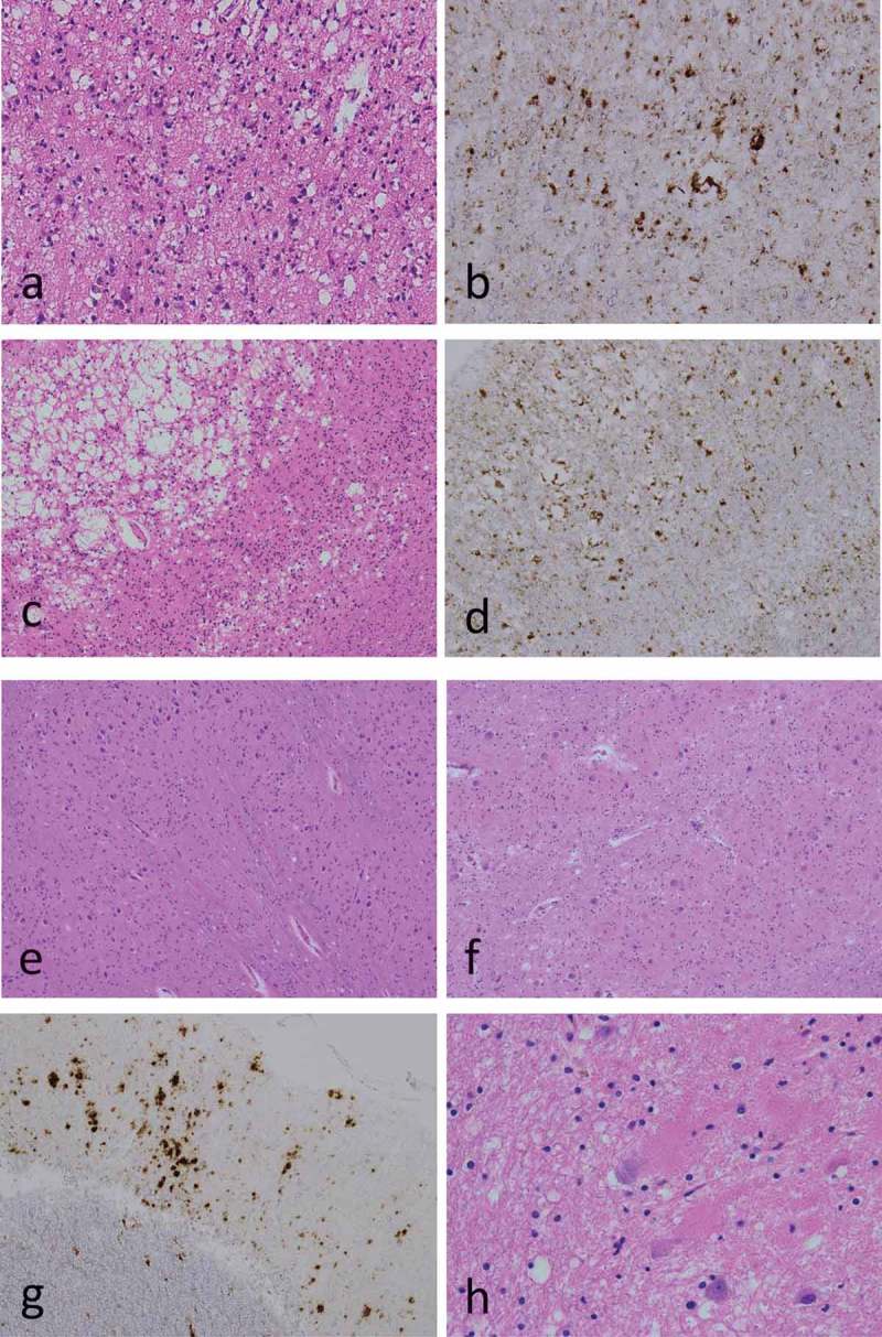
Figure 2.

Microscopic findings.
Hematoxylin-eosin stains (a, c, e, f, h); anti-PrP immunostains using 3F4 antibodies (b, d, g); middle temporal gyrus (a, b); primary visual cortex (striate area) (c, d); inferior olivary nuclei (e); cerebellar cortex (f, g); cerebellar dentate nuclei (h). Neuropathological examination revealed large confluent vacuole type spongiform changes in the neocortices (a, c); however, no spongiform changes were found in the inferior olivary nuclei (e). PrP immunostaining showed PrP deposits around the vacuoles (perivacuole-type), and rough plaque-type PrP deposits (coarse-type) in mainly in the neocortices (b, d, g). Grumose degeneration was found in the cerebellar dentate nuclei (h).
