
Figure 2.
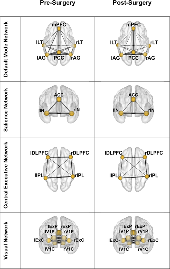
Surgery group mean node connectivity changes from pre to post time points. Line thickness between nodes is weighted by node-to-node correlation. Lowercase “r” and “l” denote right and left brain hemispheres, respectively. Node abbreviation is as follows:
DMN: mPFC – medial prefrontal cortex; LT – lateral temporal; AG – angular gyrus; PCC – posterior cingulate cortex; SN: ACC – anterior cingulate cortex; IN – insula; CEN: DLPFC – dorsolateral prefrontal cortex; IPL – inferior parietal lobe; VN: ExP – extrastriate peripheral fields; V1P – peripheral visual cortex; ExC – extrastriate central fields; V1C – central visual cortex