
Figure 3.
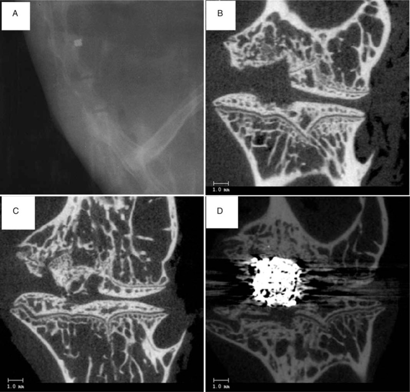
Imaging of operative lumbar spine segments of the New Zealand rabbits. (A) Post-operative lateral radiograph showing a non-radiolucent tantalum implant was implanted into the L3–L4 intervertebral space. (B) Micro-computed tomography (micro-CT) image of discectomy only space (control group) showing the appearance of the defect after discectomy in the intervertebral space. (C) Micro-CT image showing discectomy with autologous bone implanted space (autograft group). (D) Micro-CT image showing discectomy with porous tantalum implanted space (tantalum group).