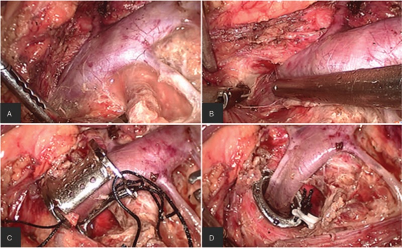
Figure 3.

The stent was placed and fixed step by step. The LRV, the LACV, and the left reproductive vein were exposed (A); the compressed LRV between SMA and aortaventralis was freed by cutting off the surrounding fibrous tissue, and the IVC was also exposed (B); the stent was placed around the LRV (C); the stent was pushed between the SMA and aortaventralis, and the dialated LRV collapsed (D). IVC: Inferior vena cava; LACV: Left adrenal central vein; LRV: Left renal vein; SMA: Superior mesenteric aorta.
