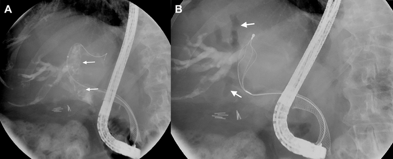
Fig. 5. Cholangiographic images of hemobilia.

(A) Intraductal lucencies (arrows) suggestive of occlusive clot formation are seen fluoroscopically. A self-expanding metallic stent is also noted in the biliary tree. (B) A flower basket is used to extract clot from the biliary tree. Resultant air cholangiograms are seen, with arrows denoting pneumobilia.
