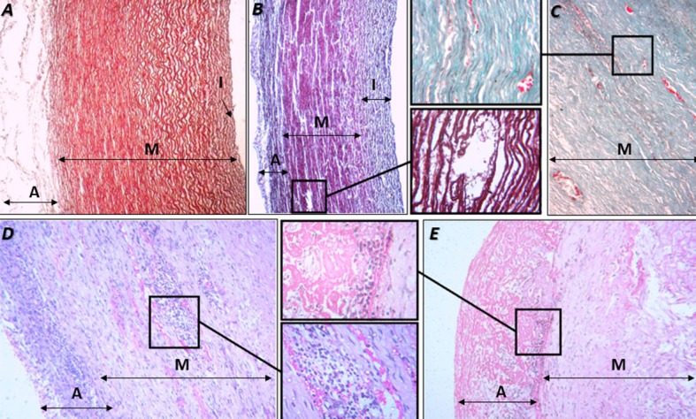
Figure 1. Photomicrograph of the aorta (partial). A) non-surgically manipulated area. Weigert-van Gieson stain (40x). B-E) surgically manipulated area, as follows: B) artery with intimal thickening. Weigert-van Gieson stain (40x). Detail: tissue disruption in the media layer (400x). C) media layer. Masson’s trichrome stain (100x). Detail: vasa vasorum and neoangiogenesis (400x). D) Adventitia and media layers. Hematoxylin and eosin (HE) stain (100x). Detail: inflammatory infiltrate and neoangiogenesis in the media layer (400x). E) Adventitia and media layers. HE stain (100x). Detail: inflammatory infiltrate and disorganization of the adventitia (400x). A = adventitia; I = intima; M = media.

