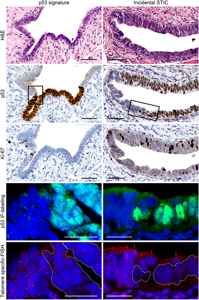
Figure 1. Representative images of a p53 signature and an incidental STIC without concurrent HGSC.

In H&E staining, the p53 signature is a cytologically benign secretory cell-rich segment. The lesion shows diffuse p53 staining pattern and a low Ki-67 labeling index by immunohistochemistry. STIC exhibits nuclear stratification, enlargement, and prominent nucleoli. It shows diffuse p53 expression and a high Ki-67 labeling index. p53 IF-labeling and telomere-specific FISH images represent higher magnification of the area marked by rectangle in p53 staining. The p53 IF-labeling shows p53-positive and -negative regions of the sections. In telomere-specific FISH, the telomere signals are weaker in the p53-positive cells within the p53 signature and the STIC lesion (white dotted lines) compared with the p53-negative, adjacent NFT epithelium (red arrows). Nuclei are stained with DAPI (blue), p53 is labeled with green nuclear fluorescence, and telomeres are hybridized with a Cy3-labeled telomere-specific FISH probe (small red dots in nuclei).
Scale bars: black, 50 μm; white, 20 μm.
H&E, hematoxylin and eosin; IF, immunofluorescence; FISH, fluorescence in situ hybridization; STIC, serous tubal intraepithelial carcinoma; HGSC, high-grade serous carcinoma; NFT, normal-appearing fallopian tube.
