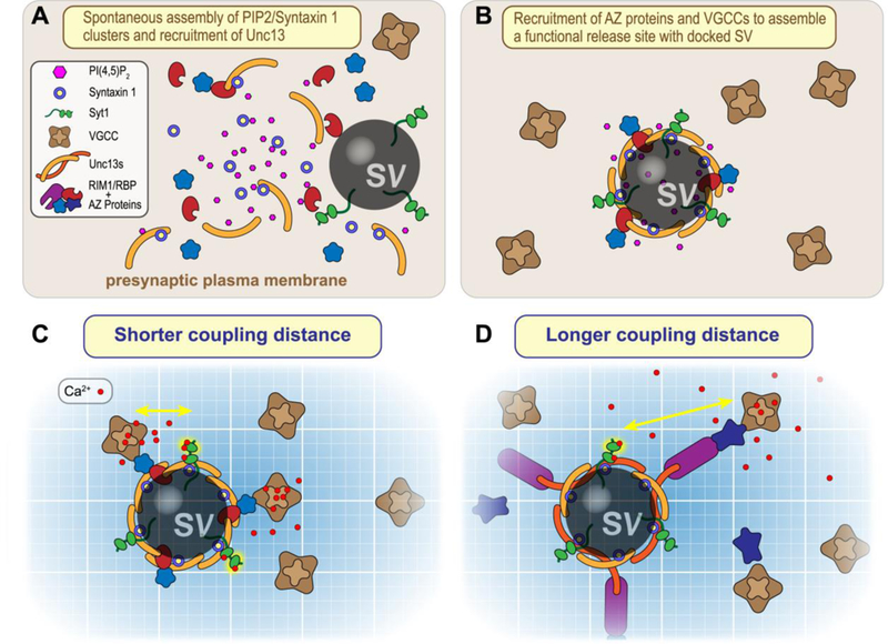
Figure 3.

Nucleation of release site assembly by Unc13, PIP2>, and Syntaxin. A. Plasma membrane Syntaxin 1 (blue/yellow) and PI(4,5)P2 (pink) tend to coalesce into a nanodomain that can recruit Unc13 (orange) via PIP2 interactions with its C2B domain as well as via SNARE binding to the MUN domain. B. Unc13 nanoassemblies recruit other AZ proteins (blue and red) to create a functional release site. C & . Distinct Unc13 isoforms together with AZ proteins couple voltage-gated calcium channels (VGCCs) at different distances from the calcium sensor Synaptotagmin 1 (Syt1, green). Calcium ions entering the presynaptic terminal indicated in red.
