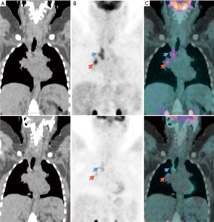
Figure 11.
18F-FDG-PET/CT images of a 29-year-old woman who was the daughter of an active TB case and diagnosed with latent tuberculosis infection. (A) Coronal CT images. (B) Coronal PET images. (C) Coronal PET/CT fused images demonstrating FDG uptake in the right paratracheal region (blue arrow) and in the right hilar region (red arrow). Top panels are the initial study, and bottom panels are the study after 3 months of treatment with isoniazid demonstrating reduction in 18F-FDG uptake. Reproduced with permission from (31). FDG, fluorodeoxyglucose; PET, positron emission tomography; CT, computed tomography.

