
Figure 4.
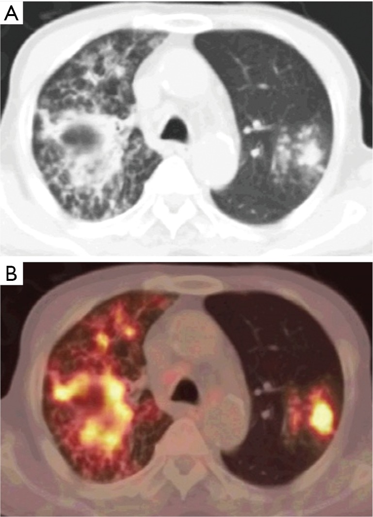
Pulmonary tuberculosis in a 61-year-old man. Axial CT (A) and axial 18F-FDG-PET/CT (B) show multiple FDG-avid pulmonary lesions. There is a consolidation with central cavitation-peripheral FDG concentration and central cold areas in the right lung suggesting an active disease. FDG, fluorodeoxyglucose; PET, positron emission tomography; CT, computed tomography.