Figure 4.
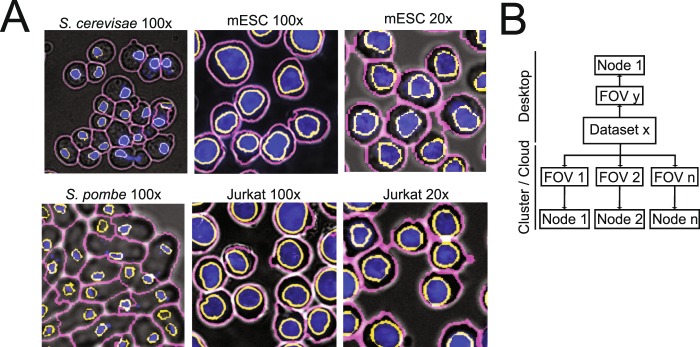
Application, implementation and performance of cell segmentation for different cell types from different organism imaged at low and high resolution. (A) Overlay of widefield (grey), DAPI (blue), nuclear (yellow) and cytoplasmic (magenta) segmented S. cerevisiae, S. pombe, mESC and Jurkat cells imaged at 100x and mESC and Jurkat cells imaged at 20x. (B) Image processing can be performed on a desktop computer or on a computing cluster for each individual image stack resulting in significant throughput. Segmenting one image takes on overage 30 minutes. For an average time course experiment consisting of 64 images of yeast cells, parallelization speeds up the image processing 64 fold.

