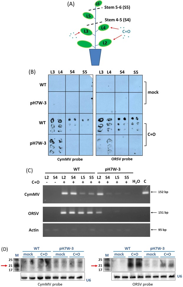
Figure 4.
CymMV and ORSV viral RNA and siRNA accumulation in WT and transgenic plants inoculated with C+O. (A) Illustrations of sampling tissues in WT and transgenic line pH7W-COCP line3 (pH7W-3) after Leaf 3 (L3) and leaf 4 (L4) co-inoculated with C+O. L3, L4, Stem between L4 and L5 (S4), Stem between L5 and L6 (S5) and systemic leaf 5 (L5) were harvested for tissue blotting (B) and RT-PCR (C) at 14 dpi. (B) The tissue blots prepared from various tissues of WT and transgenic line were hybridized with DIG-labeled CymMV- and ORSV-specific CP probes. (C) Accumulation of CymMV and ORSV viral RNA in L2, S4, S5 and L5 of WT and transgenic line by RT-PCR at 14 dpi. CymMV RNA was detected by primer pair (CymMV RdRp-F and CymMV RdRp-R) of expected size 152 bp and ORSV RNA by primer pair (ORSV RdRp-F and ORSV RdRp-R) of 151 bp. Actin was used for loading controls. (D) Detection of the vsiRNAs in mock and C+O infected IL of WT and transgenic pH7W-3 at 10 dpi by [γ-32P] CTP-labeled CymMV or ORSV CP probes. The lower panel shows hybridization of U6 probes as equal loading control. The positions of 21 nt RNA is indicated by arrows. M: marker; mock: water inoculation; C+O: inoculated with CymMV and ORSV.

