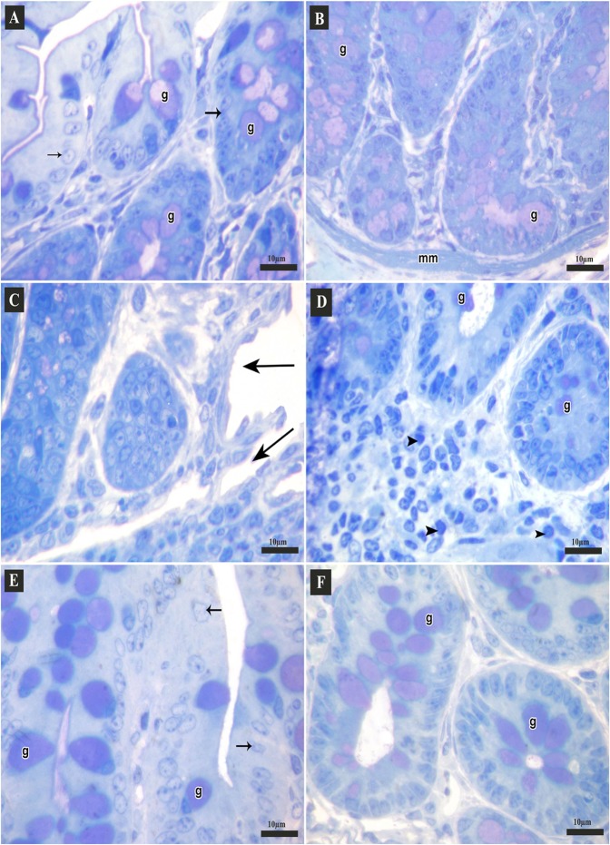
Figure 5.
Semi-thin sections of the colon of adult male mice (Toluidine blue stain, 1000x). (A,B: Control group A) Apical parts of the mucosa; columnar cells (arrows) and many goblet cells (g) that may open into the lumen. (B) Basal parts of the crypts lined with many goblet cells (g), muscularis mucosa (mm). (C,D: DSS group C) Apical parts of mucosa with mucosal ulcers (arrows). (D) Basal parts of the crypts; few goblet cells (g), diffuse inflammatory cell infiltrations (arrow heads). (E,F: DSS+ AuNPs group E) Apical parts of the mucosa, columnar cells (arrows), many goblet cells (g). (F) Basal parts of the crypts, many goblet cells (g).

