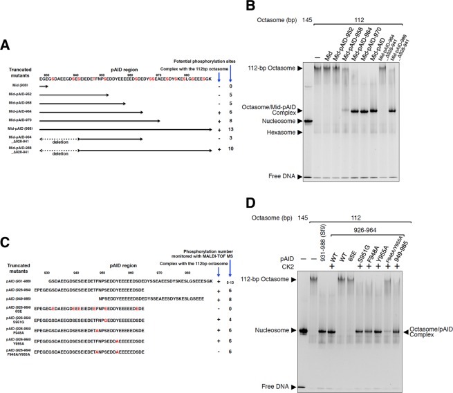
Figure 3.
Systematic analysis of the complex formation of the 112-bp octasome with the mutants of Mid-pAID and pAID. (A) Schematic drawing of the truncated mutants of Mid-pAID. These mutants were used in (B). The amino acid sequence of SPT16 (residues 928–988) is presented. The potential phosphorylation sites are colored red. (B) EMSAs show the complexes of the 112-bp octasome with the truncated mutants of Mid-pAID, detected by SYBR Gold nucleic acid gel stain. Experiments were repeated at least three times. (C) Schematic drawing of the pAID mutants. These mutants were used in (D). The amino acid sequence of SPT16 (residues 926–988) is presented. The mutated residues are colored red. (D) EMSAs show the complexes of the 112-bp octasome with the mutants of pAID, detected by SYBR Gold nucleic acid gel stain. Experiments were repeated at least three.

