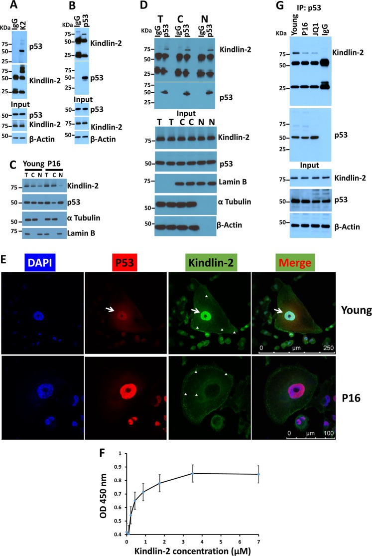
Fig. 4. Kindlin-2 interacts with p53 in both the cytoplasm and the nucleus of cancer cells.
a, b Protein lysates prepared from early passage (young) MDA-MB-231 cells was used for immunoprecipitation with mouse anti-Kindlin-2 antibody or control mouse IgG a; and mouse anti-p53 antibody or control mouse IgG b and subjected to immunoblotting analysis with rabbit anti-p53 antibody (a, upper panel) or mouse anti-Kindlin-2 antibody (b, upper panel). In control blots, cell lysates were also immunoblotted with mouse anti-Kindlin-2 and rabbit anti-p53 antibodies to show the presence of equal amounts of these proteins in the cell lysates (input panels). β-Actin is a loading control. c Total, nuclear and cytoplasmic fractions of protein lysates from early passage (young) and senescent (P16) MDA-MB-231 cells were subjected to immunoblotting with the indicated antibodies. d Total, nuclear and cytoplasmic fractions of protein lysates from early passage (young) MDA-MB-231 cells were used for immunoprecipitation with mouse anti-p53 antibody or control mouse IgG, and subjected to immunoblotting analysis with mouse anti-Kindlin-2 antibody (upper panel) or rabbit anti-p53 antibody (upper panel). In control blots (input panels), protein lysates fractions were also immunoblotted with mouse anti-Kindlin-2 and rabbit anti-p53 antibodies to show the presence of equal amounts of these proteins in the lysates as well as the specificity for the nuclear (Lamin B) and the cytoplasmic (α Tubulin). β-Actin is a loading control. e Confocal microscopy images of immunofluorescence staining of early passage (young) or senescent (P16) MDA-MB-231 cells that were stained for p53 (Red) or Kindlin-2 (Green). Nuclei were counterstained with DAPI. White arrows point to the co-localization of Kindlin-2 and p53 in the nucleus of young but not senescent cells in the merged image. The white arrowheads point to the localization of Kindlin-2 to focal adhesions in both young and senescent cells. Higher magnifications of other representative images are shown in Supplemental Fig. 2. f Interaction of Kindlin-2-GST with purified p53. The binding isotherms of increasing concentrations of GST-tagged WT Kindlin-2 to the wells of microtiter plates coated with p53. The data are expressed as means ± SEM of triplicates of two representative experiments. g Protein lysates prepared from early passage (young), passage 16 (P16) or JQ-1 treated (1 mM for 2 days) MDA-MB-231 cells was used for immunoprecipitation with mouse anti-p53 antibody or control mouse IgG and subjected to immunoblotting analysis with rabbit anti-Kindlin-2 antibody or rabbit anti-p53 antibody (upper panels). In control blots (input panels), cell lysates were also immunoblotted with mouse anti-Kindlin-2 and rabbit anti-p53 antibodies to show the presence of equal amounts of these proteins in the cell lysates. β-Actin is a loading control

