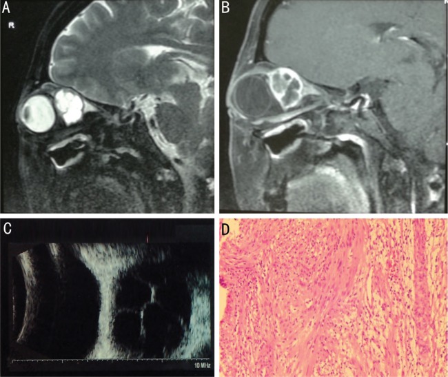
Figure 1. A 47-year-old woman experienced a gradually progressive proptosis with visual deterioration of the right eye and diplopia for one year.
A, B: T2-weighted sagittal MRI and enhanced T1-weighted MRI showed a well-defined multicystic-like lesion mass; C: B-mode ultrasound examination revealed liquid dark areas with central septa; D: The tumor was histologically confirmed as neurilemmoma with both Antoni A and Antoni B portions.

