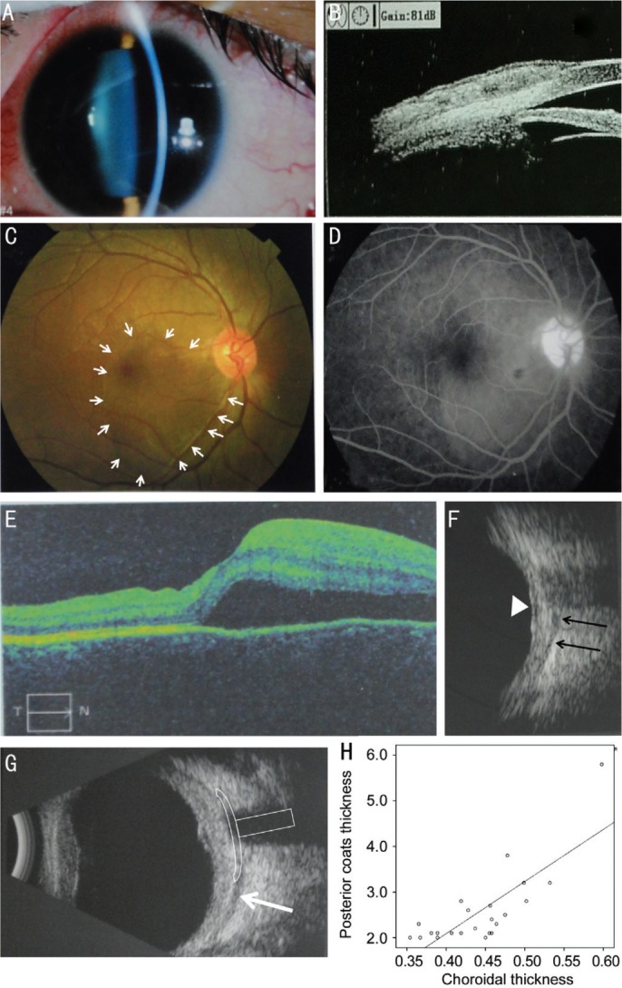
Figure 2. Clinical findings of posterior scleritis with serous retinal detachment.

A: Anterior segment examination showed conjunctival congestion and anterior scleritis with tenderness in the upper part of the sclera; B: UBM examination detected swelling and hyperemia in the superficial fasciitis and sclera at the 12 o'clock; C: Posterior segment examination revealed retinal detachment at the macula (white arrow); D: FFA illuminated the serous retinal detachment at the macula but no fluorescein leakage was detected; E: The OCT scanning confirmed serous retinal detachment at the macula; F: B-scan ultrasonography confirmed scleral thickening (black arrows indicate the posterior boundary of the sclera and the retinal detachment is indicated by the white triangle); G: B-scan ultrasonography showing diffuse thickening of the posterior coats of the eye (3.6 mm) together with fluid in the Tenon's capsule (white arrow). The “T” sign is indicated by the white hollow shape. H: The increases in the PCT correlated positively with the increasing CT (r=0.783, P<0.001).
