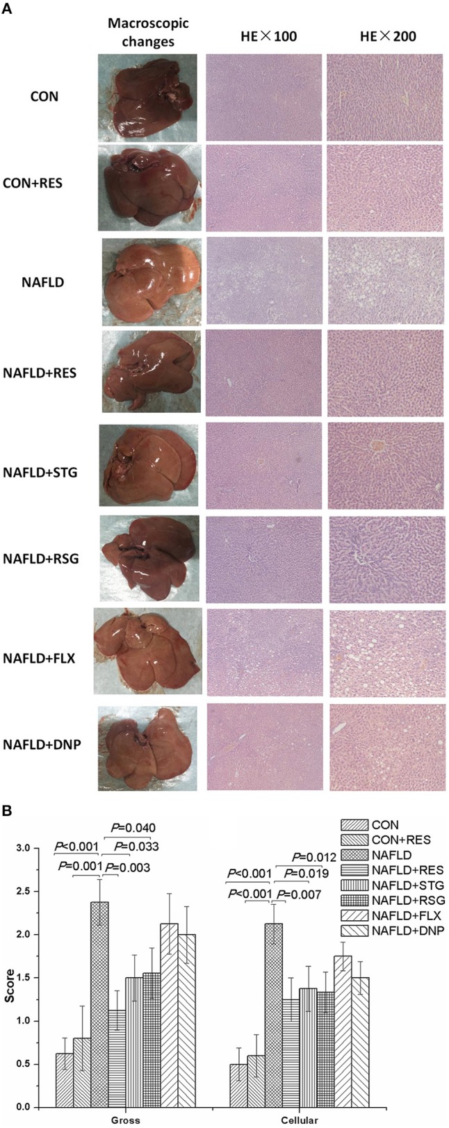
Figure 3.

Effect of RES on the macroscopic and histological liver changes in NAFLD rats. The livers of the NAFLD rats appeared yellow or pale, greasy, and brittle. Inflammatory cells and numerous lipid droplets were detected in the livers of NAFLD rats via HE staining (A). The gross and cellular scores of the liver histological changes in the NAFLD model rats were significantly increased (B). Compared with those in the NAFLD group, the color and texture of the livers of the RES-treated rats were closer to normal, and the gross and cellular scores were significantly decreased.
