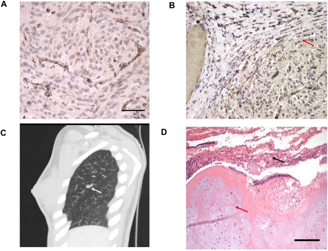
Figure 3.
Figure (A) and (B) shows loss of SDHB protein expression on immunohistochemical analysis of the primary wtGIST tumour in case #001 and #003 respectively. In Figure (B) SDHB expression is preserved in adjacent normal tissue as highlighted by the red arrow. Figure (C) shows a pulmonary chondroma in case #021 as demonstrated by the white arrow and Figure (D) demonstrates the histology of a pulmonary chondroma from case #004, with evidence of normal collapsed lung tissue illustrated by the black arrow and chondrocytes in the tumor marked by the red arrow.

