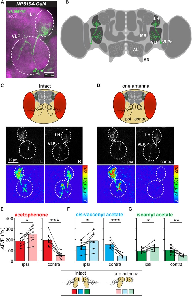
FIGURE 1.

Asymmetric odor stimulation induces contralateral suppression. (A) Confocal z-projections of VLPn labeled by NP5194-Gal4 in an adult female brain. Labeling of GFP (green) and the neuropil marker nc82 (magenta) are shown. Dashed circles represent the lateral horn (LH) and ventrolateral protocerebrum (VLP). Scale bar = 20 μm. (B) Schematic illustration of the Drosophila brain showing the innervation pattern of the VLPn in the LH and the VLP. Mushroom body (MB), antennal lobe (AL), and antennal nerve (AN) are shown for orientation. (C,D) Upper panel: Schematic head of Drosophila with intact antennae (C) and after removal of the third antennal segment of one antenna (D). Middle panel: Gray-scale image represents the VLPn structure expressing GCaMP6f. Dashed circles indicate the LH and the VLP in the left (L) and right (R) brain hemispheres. Scale bar = 50 μm. Lower panel: false-color coded images showing odor-evoked responses from a representative animal before (C) and after (D) antennal removal. Dashed circles represent the LH and VLP. (E–G) Calcium signals obtained with two-photon imaging from flies bearing UAS-GCaMP6f under control of NP5194-Gal4 from the ipsi- and contralateral sides (to the intact antenna) before and after antennal removal to stimulation with acetophenone (E), cis-vaccenyl acetate (F), and isoamyl acetate (G). Dots and lines represent individual flies, bars represent the mean (n = 8; *p < 0.05; ∗∗p < 0.01; ∗∗∗p < 0.001, paired t-test).
