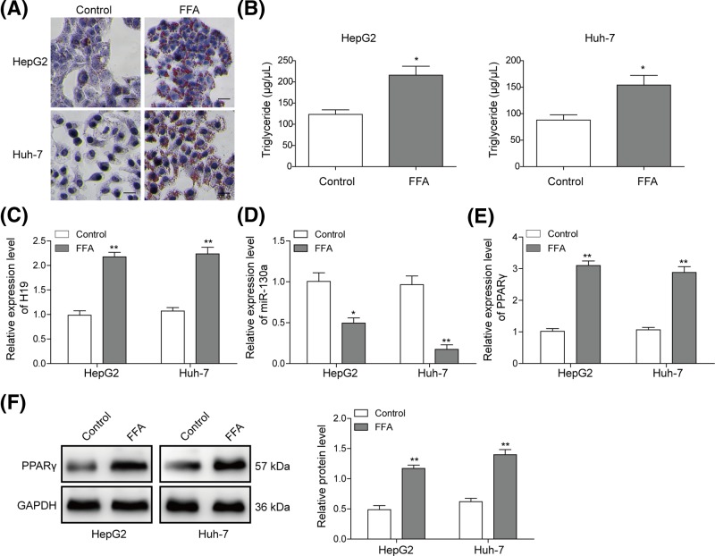
Figure 2. FFA induced hepatocyte lipid degeneration and increased H19 expression in hepatocytes.
(A) Oil-Red O staining for lipid droplet in HepG2 and Huh-7 cells treated with FFA. (B) TG levels were detected by ELISA assay in HepG2 or Huh-7 cells treated with FFA. Relative expression level of H19 (C), miR-130a (D) and PPARγ (E) was detected by qRT-PCR in HepG2 or Huh-7 cells treated with FFA. (F) Protein level of PPARγ was evaluated by Western blotting in HepG2 or Huh-7 cells treated with FFA. GAPDH was used for normalization. All the results were shown as mean ± SD (n=3), which were three separate experiments performed in triplicate. *P<0.05 and **P< 0.01.

