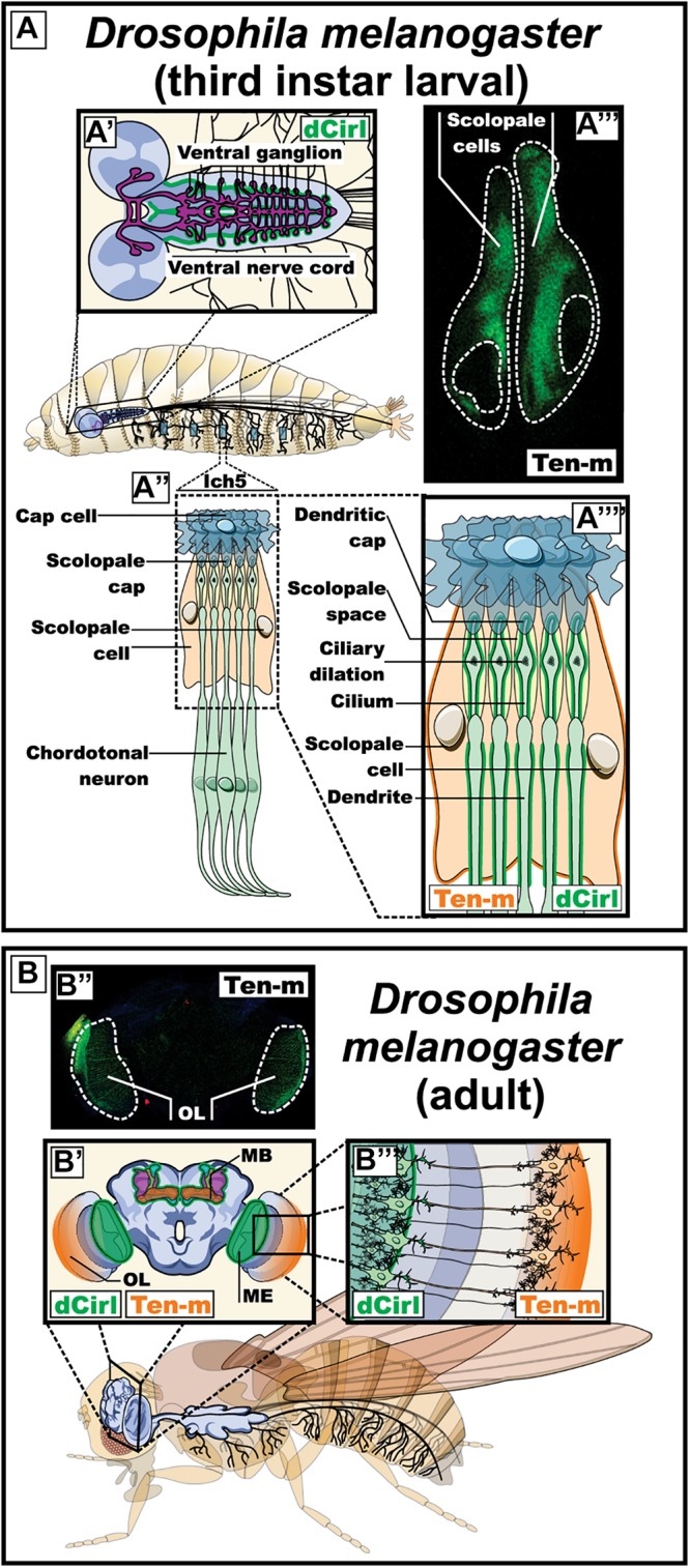
FIGURE 5.

Expression pattern of dCirl and its possible ligand Ten-m in Drosophila melanogaster (fruit fly). (A) Expression of dCirl and Ten-m during the larval stage. A′) Schematic representation of the central nervous system indicating the reported expression of dCirl in the ganglion of the ventral nerve cord (in green) (Scholz et al., 2015). A″, A″″) Magnified representation of the sensory neurons in the pentascolopidial chordotonal organs (lch5) showing the reported expression pattern of dCirl in the dendritic membrane and the single cilium of chordotonal (ChO) neurons (in green) (Scholz et al., 2015). A″′) Fluorescence microscopy image showing Ten-m promoter-driven expression of YFP in the scolopale cells of the lch5 (in green). (B) Expression of dCirl and Ten-m during the adult stage. B′) Coronal view representation of the adult brain depicting dCirl expression in the medulla of the optic system and in the mushroom bodies (in green) (Gehring, 2014). B″) Fluorescence microscopy image showing Ten-m promoter-driven expression of YFP in the optic lobe (in green). B″′) Schematic representation of contacting photoreceptor neurons in the eye expressing Ten-m (in orange) and dCirl (in green) (Gehring, 2014). OL, optic lobe; MB, mushroom body; ME, medulla of the optic system.
